EEG/Neurostimulation Core
The EEGC consists of several laboratories for the conduct of high-density EEG neuroimaging experiments, and possesses the capability of
non-invasive neurostimulation. The overarching enterprise of the CVCN is to facilitate the ability of our researchers to conduct experiments
that shed light on the relationship (causal or correlational) between nervous system activity and human sensation, perception, cognition
and action. The history of vision science has proven that indirect measures of neural function, such as sensory thresholds or perceptual
matches, are a remarkably powerful tool for the “psychoanatomical” dissection of nervous system structure and function, but recent advances
in neuroimaging technology have resulted in the development of non-invasive methods allowing the direct observation of ongoing brain
psychophysical experimentation and indirect inferences can be supplemented and augmented by parallel experiments that directly reveal
behavioral tasks, the ability to draw causal inferences regarding the neural substrates of perceptual and cognitive functions is vastly increased.
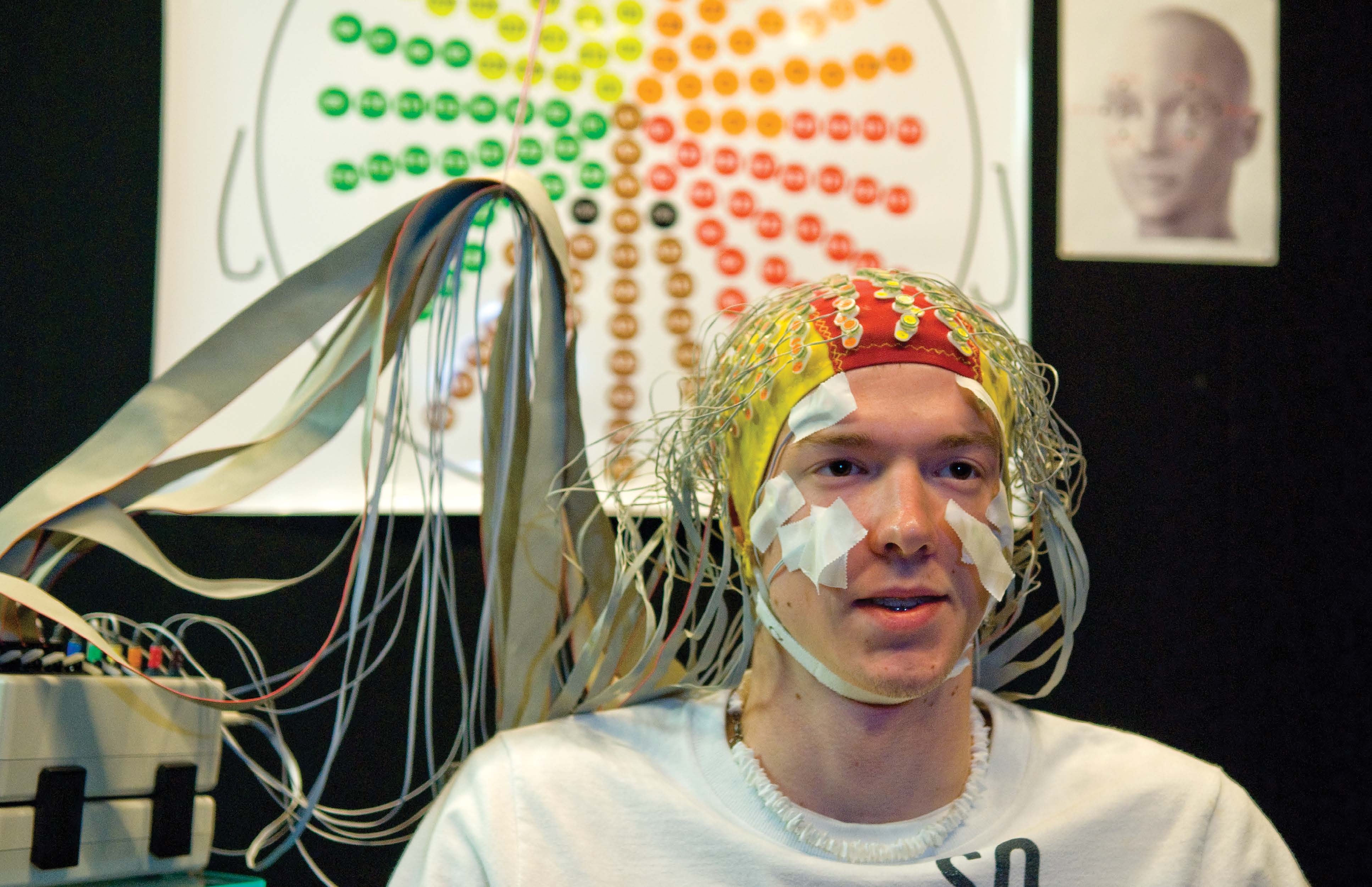
EEG Recording Instrumentation:
(2) BioSemi recording suites
-
- Active Two 8-channel amplifier/converter module
-
- 64- and 160-channel active electrode caps
-
- LabView recording and visualization software
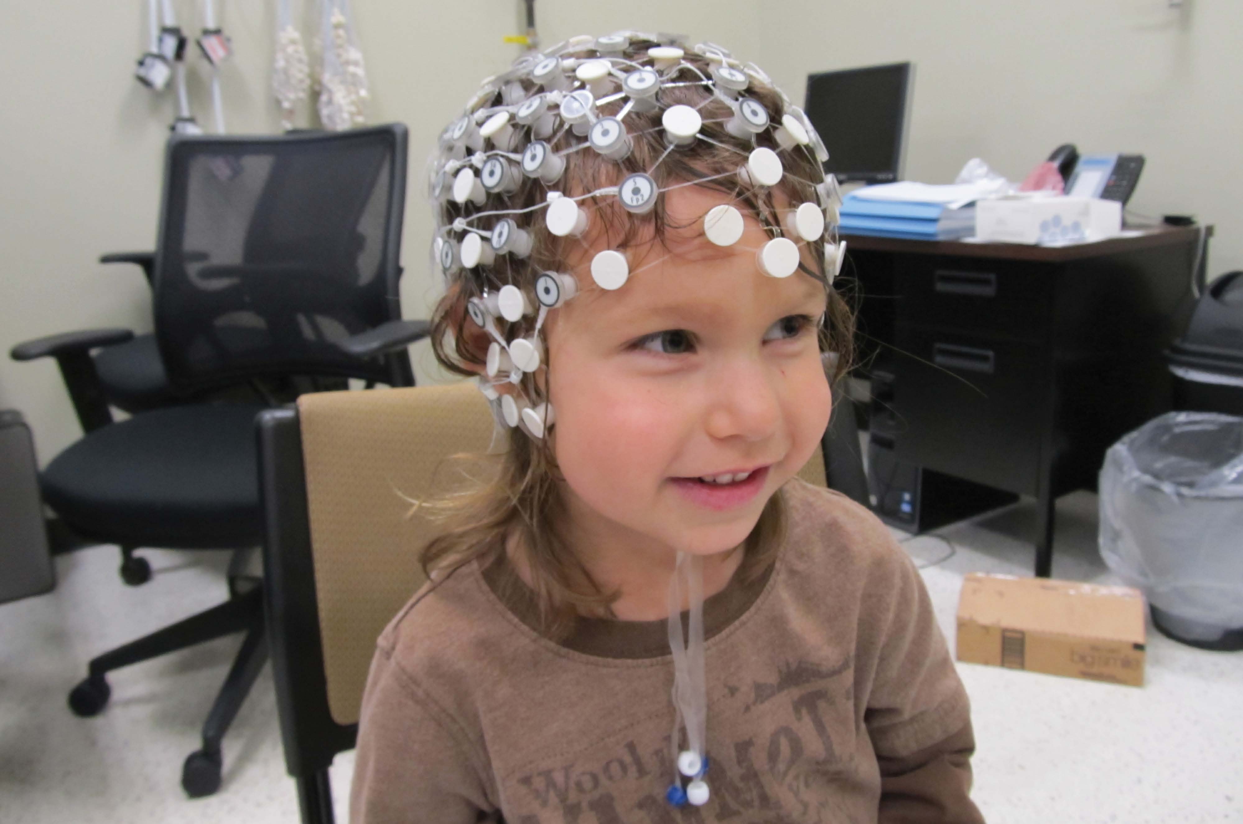
(1) Electrical Geodesics recording suite
-
- NetAmps multi-channel amplifier
-
- 64 channel Hydrocell Geodesic Sensor Nets
-
- NetStation software for data acquisition and analysis
-
- GeoSource software for source localization
(1) Wearable Sensing (Quasar) EEG recording suited
|
|
|
EEG Recording Chambers:
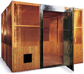 The BioSemi and EGI EEG recording equipment is installed in
(2) 8’ x 10’ x 8.75’ radio frequency (RF) shielded enclosures.
RF-shielding is required because scalp-recorded electrical signals
generated by brain activity are measured in microvolts, and these tiny potentials must be highly amplified for recording and analysis. These fields, arise from AC current in electrical wiring, and/or from the power supplies of laboratory equipment. The RF-shielded
recording chambers (Lindgren, Inc.) installed during Phase I of the COBRE reduces magnetic field strength at 60 Hz by 30 dB, and by 100dB at 14 kHz. The reduction of EM contaminants ensures that
our electrophysiological experiments are conducted in an electromagnetically and acoustically quiet environment.
The BioSemi and EGI EEG recording equipment is installed in
(2) 8’ x 10’ x 8.75’ radio frequency (RF) shielded enclosures.
RF-shielding is required because scalp-recorded electrical signals
generated by brain activity are measured in microvolts, and these tiny potentials must be highly amplified for recording and analysis. These fields, arise from AC current in electrical wiring, and/or from the power supplies of laboratory equipment. The RF-shielded
recording chambers (Lindgren, Inc.) installed during Phase I of the COBRE reduces magnetic field strength at 60 Hz by 30 dB, and by 100dB at 14 kHz. The reduction of EM contaminants ensures that
our electrophysiological experiments are conducted in an electromagnetically and acoustically quiet environment.
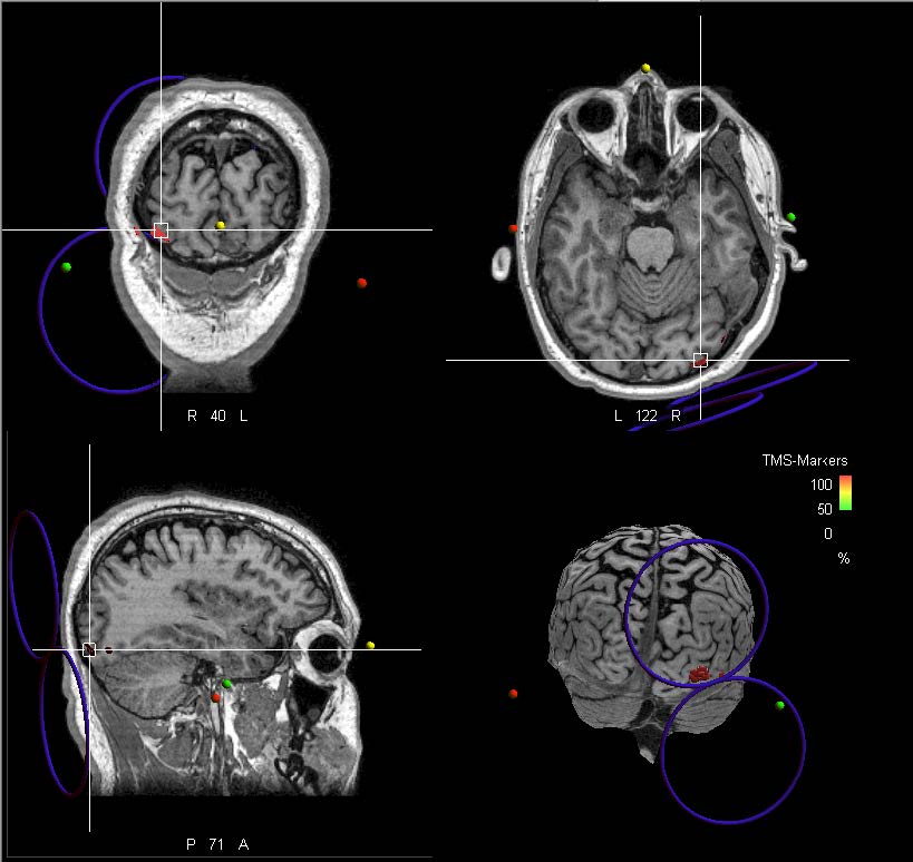
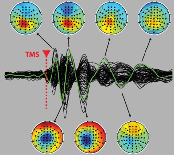
EEG/Neurostimulation Instrumentation:
(1) ANT EEG recording suite
-
- ASALab Transcranial Magnetic Stimulation (TMS)-compatible amplifier
-
- (3) WaveGuard 64 channel shielded electrode caps
-
- (3) EasyCap 64 channel TMS-compatible electrode caps
-
- ASALab recording and visualization software
(1) Magstim Super Rapid2 TMS stimulator
-
- Rapid2 capacitor and system mainframe
-
- 70 mm air-cooled TMS coil
-
- 70 mm standard coil
-
- Two-channel EMG preamplifier pod
(1) Visor2 3D TMS Neuronavigation system
-
- Visor2 software
-
- NDI Vicra tripod mounted infrared camera
-
- Xensor 3D electrode digitizer module
|
|

|
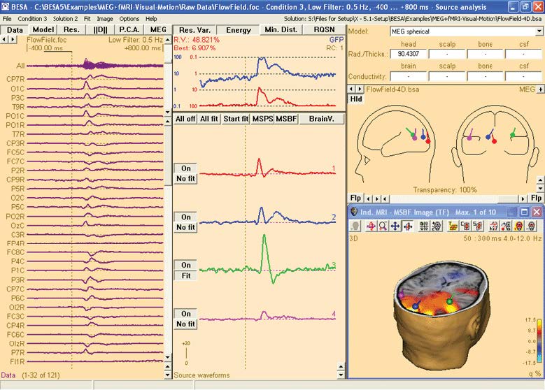
Data Storage and Analysis Infrastructure:
(3) Multiuser data analysis workstations
-
- Dell Precision 7600 multi-core desktop computer
-
- 48 Gb RAM
-
- BESA proprietary software
-
- Matlab and compatible open-source toolboxes
(1) Dell PowerEdge 720 Server
- 20 processing cores
-
- 148 Gb RAM, ~10 Tb storage
-
- Matlab and compatible open-source toolboxes
|
|
|