Title
Sunflower Disease Diagnostic Series
(PP1727, Reviewed Jan. 2023)File
Publication File:
PP1727 Sunflower Disease Diagnostic Series
Summary
This series aids in disease identification.
Availability
Availability:
Web only
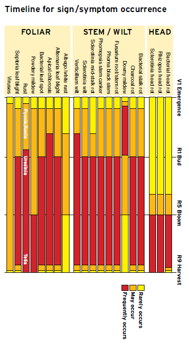
Publication Sections
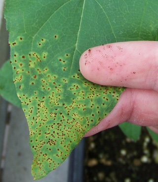
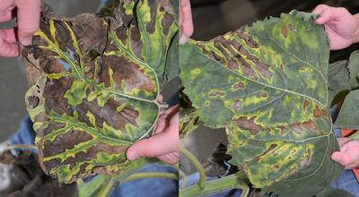
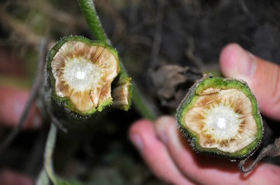
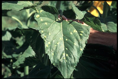
Bacterial head rot
Pectobacterium carotovorum, subsp. carotovorum and P. atrosepticum
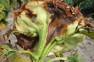
FIGURE 1 – Watery lesions forming on heads as a result of infection through wounds
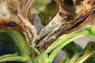
FIGURE 2 – Slimy masses of bacterial growth within infected head tissues
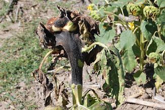
FIGURE 3 – Affected tissues dry out and turn black after a period of warm, dry weather
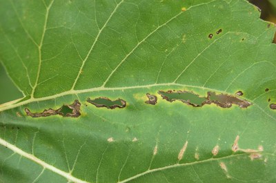
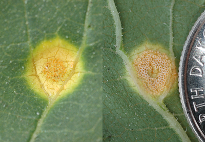
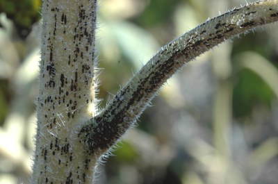
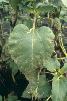
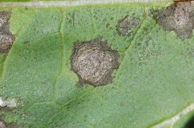
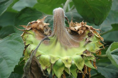
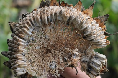
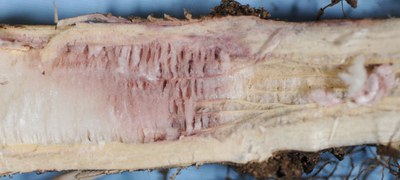
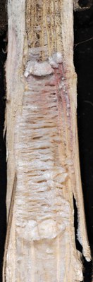
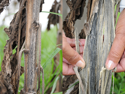
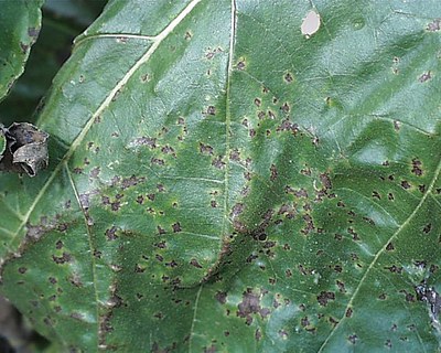
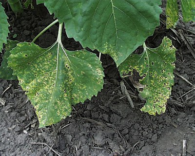
Rhizopus head rot
R. stolonifer, R. oryzae (syn. R. arrhizus) and R. microsporus

FIGURE 1 – Note wound from hail stone with subsequent development of watery, soft rot
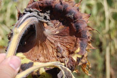
FIGURE 2 – Rotted area of head drying, shriveling and beginning to shred
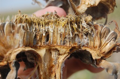
FIGURE 3 – Grayish fungal strands growing through head;

reproductive structures

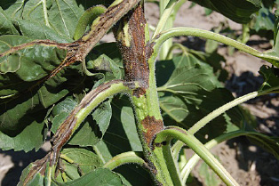
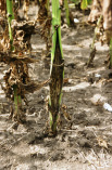
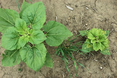
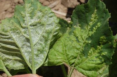
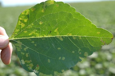
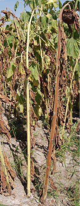
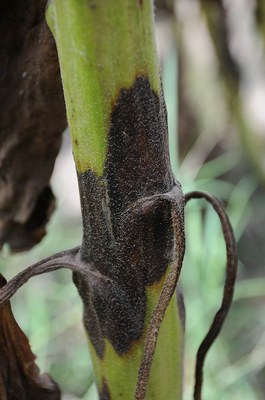


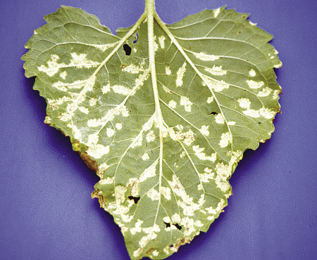
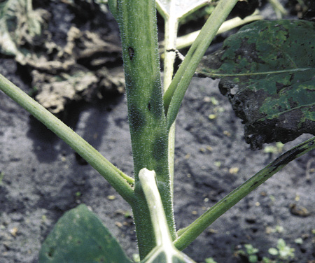
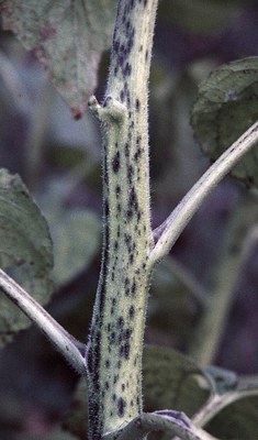
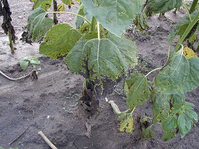
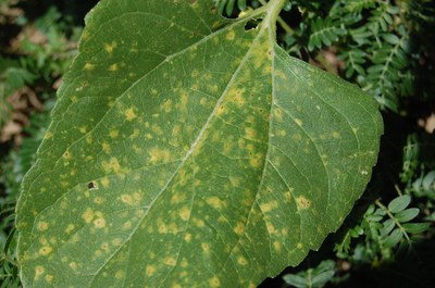
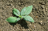
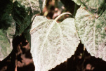
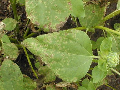
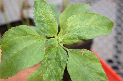
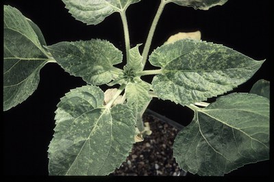
Apical chlorosis
Pseudomonas syringae pv. tagetis
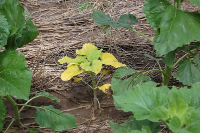
Figure 1 - Young plant infected systemically; note bright yellow chlorosis and stunting
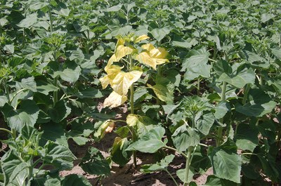
FIGURE 2 – Plant nearing bud formation (R1) exhibiting systemic chlorosis symptoms
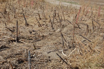
FIGURE 3 – Distribution of apical chlorosis corresponding to low areas of water accumulation in field