The culturable ocular bacterial microbiota of beef cattle and its commensal members that can inhibit pinkeye-associated pathogens
(AS2100-11, December 2023)The objective of this study was to characterize the culturable fraction of the bacterial microbiota residing in the bovine eye and to investigate whether commensal members of this community could inhibit the pinkeye-associated pathogens Moraxella bovis and Moraxella bovoculi. Results indicate that the bovine eye harbors a relatively diverse culturable bacterial community, and some of these commensal species can inhibit pinkeye pathogens, suggesting the possibility to develop bacterial therapeutics based on these commensal isolates to mitigate pinkeye infections in cattle in place of antibiotics.
Procedures
Ocular swabs from the cornea and conjunctiva of cattle exhibiting IBK symptoms (n = 35) as well as control swabs from healthy animals (n = 29) were collected from multiple herds across North Dakota as well as from the NDSU Beef Cattle Research and Teaching Center and from the NDSU Veterinary Diagnostic Laboratory (Table 1). Ocular swabs were collected using Puritan Opti-Swabs with the Liquid Amies Collection and Transport System (Puritan, Guilford, ME) and were stored on ice for transport to the lab. Once in the lab, samples were aliquoted and spread on up to five types of agar plates (Blood, Columbia Blood, De Man, Rogosa and Sharpe (MRS), Wilkins-Chalgren, and Multi-slice agar), all with various growth mediums to support the growth of a wide range of microorganisms. Plated samples were incubated both aerobically and anaerobically for 24 – 48 h. Bacterial colony growth was quantified, and unique colonies were sub-streaked onto fresh media and incubated for 24 h. Isolated bacteria were then cryopreserved (n = 658). Genomic DNA was extracted from a subset of preserved bacterial isolates (n = 351) and used for taxonomic identification by the near-full length 16S rRNA gene sequencing. Of the 351 identified isolates, 53 were tested for growth inhibitory effects against M. bovis and M. bovoculi using the agar slab method as described previously (Amat et al., 2019). Following the agar slab experiments, a selection of candidate bacteria that exhibited relatively strong inhibition of Moraxella growth were used to evaluate changes to cell morphology of Moraxella by inoculating Moraxella into the cell free supernatant of the candidate bacteria, incubating for 14 h, and observing changes using scanning electron microscopy (SEM) as described previously (Amat et al., 2019).
Table 1: Number of swabs collected and total number of isolated bacteria that were cryopreserved.
| Sources | 4 ND Veterinary Clinics, NDSU Beef Herd, NDSU VDL | No. of Summer Swabs | 43 | |||||||
| No. of Winter Swabs | 21 | |||||||||
| Swab Type | Total Number of Swabs | Aerobic Isolates | Anaerobic Isolates | Total Isolate Number | ||||||
| MP | CB | Blood | MRS | MP | Blood | WC | CB | |||
| Control | 29 | 30 | 61 | 30 | 43 | 14 | 11 | 74 | 18 | 281 |
| Pinkeye | 35 | 41 | 37 | 108 | 67 | 12 | 10 | 80 | 22 | 377 |
| Subtotal | 64 | 71 | 98 | 138 | 110 | 26 | 21 | 154 | 40 | 658 |
Results and Discussion
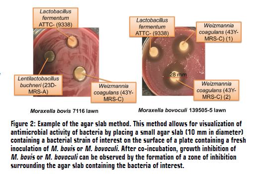
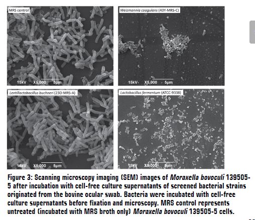
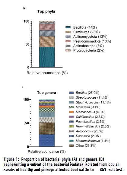
The 351 bacterial isolates identified using near-full length 16s rRNA gene sequencing were represented by 6 different bacterial phyla and 61 different bacterial genera. Bacterial phyla included Bacillota (44%), Firmicutes (23%), Actinomycetota (13%), Pseudomonadota (13%), Actinobacteria (5%), and Proteobacteria (2% Figure 1A). Of the 61 bacterial genera, Bacillus (26%), Streptococcus (11%), Staphylococcus (11%), Moraxella (9%), and Macrococcus (4%) were the most prevalent (Figure 1B). A total of 33 Moraxella isolates were identified, and they consisted of M. bovis and M. bovoculi. Of the 53 isolates tested for inhibition against Moraxella using the agar slab method (Figure 2), 6 isolates showed zones of inhibition ranging from an average of 13 mm to 25.7 mm (Table 2). Weizmannia coagulans (43Y MRS-C), Lactobacillus fermentum (ATTC 9338), and Paenibacillus polymyxa (42G WC-F) showed relatively strong inhibition against Moraxella, while Alkalihalobacillus rhizosphaerae (25F CB-B) and Lentilactobacillus buchneri (23D MRS-A) showed medium growth inhibition and Weissella paramesenteroides (23D MRS-F) displayed weak growth inhibition. Weizmannia coagulans (43Y MRS-C), Lactobacillus fermentum (ATTC 9338), and Lentilactobacillus buchneri (23D MRS-A) cell-free culture supernatants were incubated with M. bovis and M. bovoculi for 14 h and prepared for SEM imaging. The W. coagulans and L. fermentum isolates exhibited the greatest amount of cell damage to M. bovoculi. Complete cell lysis was observed, indicating that W. coagulans and L. fermentum effectively inhibit the growth of M. bovoculi. Lentilactobacillus buchneri exhibited only minor cell damage to M. bovoculi, with few structural damages to the M. bovoculi cells (Figure 3). When the cell-free culture supernatant of W. coagulans and L. fermentum was incubated with M. bovis, noticeable cell damage occurred, but it was not to the extent of the damage that occurred to M. bovoculi (Data not shown). Irregular cell shape and damages to the cell wall of M. bovis were observed, which indicates that W. coagulans and L. fermentum may be viable candidates for the development of bacterial therapeutics against M. bovis.
Table 2: Six bacterial isolates that exhibited antimicrobial activity against M. bovis and M. bovoculi when tested using the agar slab method.
| Isolate ID | Species | Average ZOI (mm) |
| 43Y MRS-C | Weizmannia coagulans | 25.7 |
| ATTC 9338 | Lactobacillus fermentum | 18.5 |
| 42G WC-F | Paenibacillus polymyxa | 17.2 |
| 25F CB-B | Alkalihalobacillus rhizosphaerae | 16.4 |
| 23D MRS-A | Lentilactobacillus buchneri | 14.1 |
| 23D MRS-F | Weissella paramesenteroides | 13.0 |
These results indicate that the bovine oculus harbors relatively diverse culturable bacteria. In addition, some of the commensal bacterial isolates can inhibit the growth of M. bovis and M. bovoculi, potentially through the production of antimicrobial agents that can damage the cell structure and cell morphology of the pathogens. This information adds to the current understanding of the bovine ocular microbiota and indicates that commensal bacterial species within the ocular microbiota may be able to be harnessed to combat against pinkeye pathogens and modulate ocular microbiome-mediated eye health in cattle.