Title

Estrogen’s role in the induction of parturition and fetal maturation in periparturient ewes

(AS2100-13, December 2023)
File
Publication File:
Summary

The objective of this study was to obtain a better picture of estrogen’s role in the control of parturition and fetal maturation in periparturient Rambouillet ewes through the evaluation of the relationship between systemic estradiol (E2) levels and 1) the timing of parturition, lamb’s birth weight, and lamb vigor, and 2) the uterine progestogenic responsiveness and progesterone (P4) plasma levels. Results suggest that estradiol may downregulate myometrial P4 receptor protein expression, leading to myometrial contraction-inducing parturition; however, it does not seem to affect lamb birth weight or vigor.

Other Authors

Bethania J. Dávila Ruiz1, Chutikun Kanjanaruch1, Carl R. Dahlen1, Pawel P. Borowicz1, Alan J. Conley2, and Lawrence P. Reynolds1

1Department of Animal Sciences and Center for Nutrition and Pregnancy, North Dakota State University, Fargo, ND USA 58108-6050
2Department of Population Health and Reproduction, School of Veterinary Medicine, UC-Davis, CA 95616

Availability
Availability:
Web only
Publication Sections

Summary

The incidence of livestock death due to premature birth is approximately 10% in the US, which results in a large economic loss for farmers and ranchers. Therefore, the prevention of premature birth and the understanding of underlying mechanisms of parturition and fetal maturation in preparation for birth are problems that need to be addressed. Parturition requires activation of myometrial contractility, which may be driven by a withdrawal of progesterone (P4) accompanied by a rise in estrogen levels. However, these relationships are unclear, as estradiol (E2) can induce parturition without P4 withdrawal. This experiment was designed to obtain a better picture of estrogen’s role in the control of parturition in periparturient Rambouillet ewes through the evaluation of the relationship between E2 levels and 1) the timing of parturition, lamb birth weight, and lamb vigor and, 2) uterine progestogenic responsiveness and systemic plasma P4 levels. Two experiments were conducted. In experiment 1, ewes were treated and allowed to deliver. The hours from treatment until delivery, and live lamb birth weight and vigor were recorded. In experiment 2, ewes were treated and slaughtered 26 h later for tissue collection. At slaughter, carotid blood was collected to measure hormone levels, and samples of the utero-placenta were formalin-fixed and immunofluorescently stained for P4 receptors. The same treatment was used for both experiments. The ewes were randomly assigned to either E (4 Silastic implants of 50 mg of E2; 200 mg/ewe; Exp 1 n = 5, Exp 2 n = 6) or C (4 empty implants; Exp 1 n = 5, Exp 2 n = 6) groups. All treatments began at d 139 to 142 of gestation. Results showed that in experiment 1, the hours from treatment to delivery were less in E compared with the C group (64.1 ± 75.64 vs 374.4 ± 75.64 h, P = 0.01), the live birth weight tended to be less in lambs from the E group (4.12 ± 0.23 vs 4.77 ± 0.21 kg. P = 0.07), but there was no difference between groups for lamb vigor (2 ± 0.39 vs 1 ± 0.48 [4-point scale with 0 being the best], P = 0.13). In experiment 2, E2 treatment downregulated P4 receptor protein expression in the myometrium of E vs C groups (27.02 ± 36.68 vs 42.0 ± 3.68 intensity units, P = 0.01), but there was no difference in systemic plasma levels of P4 in E vs C ( 6.50 ± 1.42 vs 8.99 ± 1.42 ng/mL, P = 0.24), while E2 concentration in systemic blood was less in C compared with E (30.61 ± 11.73 vs 149.21 ± 55.93 pg/mL, P = 0.01). These results suggest that in late pregnant ewes, E2 downregulates myometrial P4 receptors and thus progestogenic responsiveness of the myometrium which leads to activation of the myometrium, earlier delivery, and thus lower live lamb weight at delivery without a decrease in P4 systemic blood levels.

Introduction

Livestock death due to premature birth in the US is approximately 10%, resulting in a significant economic loss for farmers and ranchers. Therefore, addressing the problem of premature birth and understanding the mechanisms behind parturition and fetal maturation is crucial. Maintaining pregnancy and giving birth to offspring is a complex process involving multiple fetal and maternal hormones and biochemical factors, but steroids are the protagonist. Three key events must take place to ensure a successful birth: (1) the maturation of fetal organs in preparation for extra-uterine life, (2) the delivery of the fetus, and (3) the delivery of the placenta (Kota et al., 2013). The main reason behind the difficulty of defining the fundamental mechanisms that underly the initiation of parturition and fetal organ maturation is the variation between species and the limited number of mammalian species that have been studied regarding pregnancy steroidogenesis. Length of gestation, the number of fetuses, and the timing of parturition differ, and even though several hormones involved are well conserved, the tissues involved in hormone synthesis, the controlling mechanisms, and mechanisms of action differ widely among species (Conley et al., 2014; Rokas et al., 2020).

The delivery of the fetus is driven by myometrial contractions and cervical ripening, which is known to be triggered by a decrease in P4 (the pro-gestational hormone) and an increase in E2 levels. The first theory proposed for the parturition mechanism in sheep in 1973 by G. C. Liggins indicated that the fetal lamb could signal its own birth through the activation of the fetal hypothalamic-pituitary-adrenal (HPA) axis. This model postulated that when the fetus becomes stressed, possibly due to a lack of nutrients or space at the end of pregnancy, the HPA axis is activated, leading to the release of cortisol by the fetal adrenal cortex that will be transported to the placenta and will stimulate the steroidogenic enzymes involved in the conversion of P4 into estrogens, leading to the withdrawal of P4 and myometrial activation (Liggins et al., 1973). However, previous studies have demonstrated that the relationship between these hormones is unclear for many reasons: 1) P4 concentration is much greater than that of estrogen at late gestation (ng/ml vs pg/ml) meaning that even if some P4 were converted into E2, it still would not result in a reduction in P4 levels (Hamon et al., 1990); 2) There is a compartmental expression of steroidogenic enzymes: in humans, bovine, and ovine, the conversion of 17OHP4 to A4 in the Δ4 pathway is very inefficient (Conley et al., 2012); and 3) E2 can induce parturition independently of P4 withdrawal (Wu et al., 2004). Therefore, the objective of this study was to obtain a better picture of estrogen’s role in the control of parturition in periparturient Rambouillet ewes through the evaluation of the relationship between systemic E2 levels and 1) the timing of parturition, lamb birth weight, and lamb vigor, and 2) the uterine progestogenic responsiveness and P4 plasma levels.

Experimental Procedures

Two experiments were conducted performing the same treatment. Multiparous ewes were randomly assigned to either E (4 Silastic implants of 50 mg of E2; 200 mg/ewe; Exp 1 n = 5, Exp 2 n = 6) or C (4 empty implants; Exp 1 n = 5, Exp 2 n = 6) groups. All treatments began at d 139 to 142 of gestation. Implants were subcutaneously inserted in the axillary region of the ewe and were removed 2 days after parturition (experiment 1) or at slaughter (experiment 2).

In experiment 1, ewes were treated and their delivery time after treatment, live lamb birth weight, and vigor were recorded. Lamb vigor was assessed using a scale ranging from 0 to 4, where 0 represents extreme activity and vigor with the lamb standing on all four feet, 1 indicates high activity with the lamb standing on its back legs and knees, 2 represents moderate activity with the lamb active on its chest and holding its head up, 3 indicates weakness with the lamb lying flat but still able to hold its head up, and 4 indicates severe weakness with the lamb unable to lift its head and showing minimal movement.

In experiment 2, ewes were treated and slaughtered 26 h later for tissue collection. At slaughter, carotid blood was collected, and P4 and E2 plasma concentrations were measured by LC-MS and radioimmunoassay. Also, cross-sections of the uterus were collected, formalin-fixed and immunofluorescent stained for P4 receptors using DAPI counterstaining. Confocal imaging of myometrium was generated for image analysis of the receptor expression with Image-Pro Plus. Statistical significance (P < 0.05) was assessed using the MIXED procedure of SAS for both experiments.

Results and Discussion

In experiment 1, the hours from treatment to delivery were less in E compared with the C group (64.1 ± 75.64 vs 374.4 ± 75.64 h, P = 0.01). This indicates that the E group had an average day of gestation (DOG) at lambing of approximately d 142 ± 3, while the C group had an average DOG of approximately d 154 ± 3. In a more recent study (Davila-Ruiz, Reynolds, Conley et al., unpublished) we found with that a similar treatment with E2 on days 139 to 142 of gestation resulted in an average delivery day of 143 ± 1 compared with an average delivery day of 147 ± 1 in control ewes. The average gestation period in Rambouillet ewes typically falls around 147 days. Furthermore, lamb weight tended to be lower in the E group compared to the C group (4.12 ± 0.23 vs 4.77 ± 0.21 kg, P = 0.07). This observation can be rationalized as the induction of earlier delivery may result in slightly lower birth weights for the lambs. Also, the presence of both single and twin pregnancies included in the experiment can impact the birth weight of the lambs, as single pregnancies typically result in higher birth weights compared to twin pregnancies. No difference in lamb vigor was found between the E and C groups (2 ± 0.39 vs 1 ± 0.48, P = 0.13). Several factors may contribute to the lack of differences in lamb vigor. 1) The timing of lamb vigor assessment conducted a few hours after birth may have not captured initial differences that could have emerged in the following days. In fact, a couple of days later, two lambs in the E group experienced sudden death, while no death was reported in the lambs of the C group. 2) Despite the E group experiencing parturition around 10 days earlier than the C group, both groups were likely at a similar stage of lung maturity. This suggests that the timing of parturition alone may not determine lamb vigor.

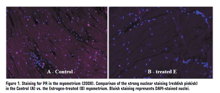
In experiment 2, a significant increase in E2 systemic levels was found in the E group compared with C group (149.21 ± 55.93 pg/mL vs 30.61 ± 11.73, P = 0.01), which suggests that E treatment effectively increased E2 levels. Estradiol has been related with the promotion of myometrial contractility and initiation of parturition. Together with the results in experiment 1, this finding suggests that a higher concentration of E2 could drive earlier delivery. A downregulation of myometrial P4 receptor protein expression was observed in the E group compared to C group (27.1 ± 36.7 vs 42.0 ± 3.68 intensity units, P = 0.01; Figure 1). This finding could have an important implication in the control of parturition, as P4 receptors play a crucial role in maintaining the inhibitory effects of P4 on uterine contractility promoting uterine quiescence during pregnancy. Therefore, the fact that the number of myometrial P4 receptors decreases with higher estrogen levels making the cells less responsive could potentially contribute to the initiation of parturition. No differences were found in P4 systemic plasma levels in E vs C (6.50 ± 1.42 vs 8.99 ± 1.42 ng/mL, P = 0.24), which together with the results in experiment 1, suggest that local changes in myometrial P4 receptors, rather than systemic P4 levels, might be more critical in regulating myometrial activation.

The findings of both experiments highlight the complex interplay between myometrial P4 receptor expression, systemic P4 and E2 concentrations, and the timing of parturition. Overall, these findings indicate that E2 concentrations have a notable effect on the timing of parturition and may contribute to slight differences in birth weight, but its impact on fetal maturation, as assessed by lamb vigor after birth, appears to be limited. Therefore, it can be concluded that E2 concentrations primarily affect the timing of parturition rather than directly influencing fetal maturation and that the downregulation of myometrial P4 receptor protein expression with the increase of E2 levels supports the important role of E2 in initiating labor. However, future research is needed to clarify the precise mechanism of parturition and fetal maturation to enhance reproductive management strategies in livestock.

Figure 1

References

Conley, A. J., and L. P. Reynolds. 2014. Steroidogenesis and the initiation of parturition. In: Reproduction in Domestic Ruminants VIII. Context Publishing, Packington UK, pp. 399-414.

Conley, A. J., C. J. Corbin, J. L. Thomas, N. A. Gee, B. L. Lasley, B. C. Moeller, S. D. Stanley, and T. Berger. 2012. Costs and consequences of cellular compartmentalization and substrate competition among human enzymes involved in androgen and estrogen synthesis. Biol. Reprod. 86:1-8.

Hamon, M. H., and R. B. Heap. 1990. Progesterone and estrogen concentrations in plasma of Barbary sheep (aoudad, Ammotragus lervia) compared with those of domestic sheep and goats during pregnancy. Reproduction 90:207–211.

Kota, S. K., K. Gayatri, S. Jammula, S. K. Kota, S. V. Krishna, L. K. Meher, and K. D. Modi. 2013. Endocrinology of Parturition. Ind. J. Endocrinol. 17:50.

Liggins, G. C., R. J. Fairclough, S. A. Grieves, J. Z. Kendall, and B. S. Knox. 1973. The mechanism of initiation of parturition in the ewe. Rec. Progr. Hormone Res. 29:111–159.

Rokas, A., S. Mesiano, O. Tamam, A. LaBella, G. Zhang, and L. Muglia. 2020. Developing a theoretical evolutionary framework to solve the mystery of parturition initiation. eLife 9 1:22.

Wu, W. X., X. H. Ma, T. Coksaygan, K. Chakrabarty, V. Collins, J. Rose, and P. W. Nathanielsz. 2004. Prostaglandin mediates premature delivery in pregnant sheep induced by estradiol at 121 days of gestational age. Endocrinology 145:1444–1452.