 |
 |
 |
 |
 |
 |
|
|
|
|
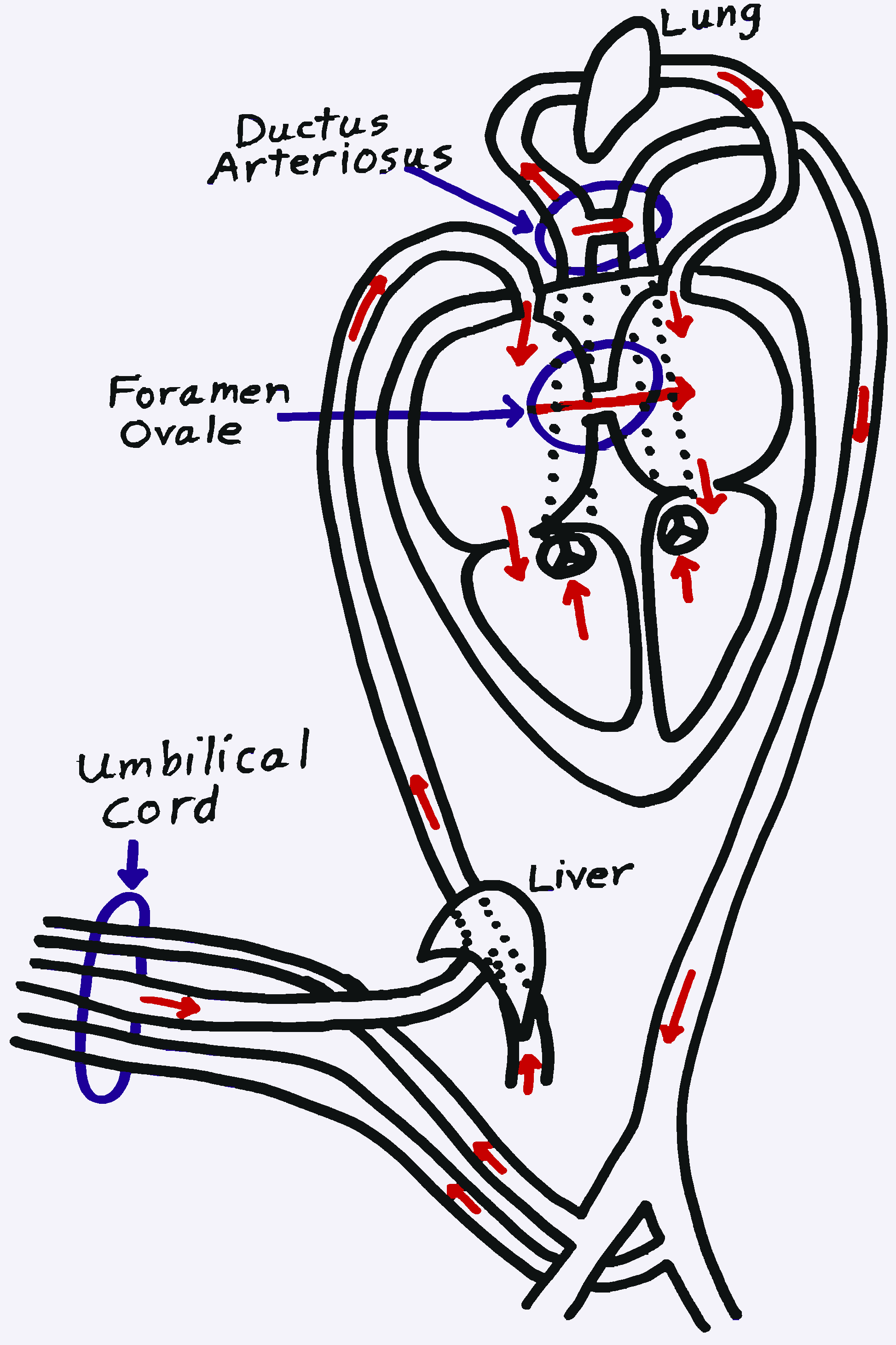 |
During its development before birth a fetus leads a parasitic existence. It is dependent on its mother for nutrients, oxygen and the elimination of wastes. The placenta is the fetus' life support system. It surrounds the fetus and attaches to the uterine wall. Its blood vessels lie very close to the uterine blood vessels, but there are no direct connections. This allows the efficient exchange of nutrients and wastes between the fetal and maternal bloodstreams. Blood vessels in the umbilical cord carry blood between the fetus and the placenta.
The nonfunctional fetal lungs and the life support functions being accomplished via the umbilical cord require some modifications in the way blood is distributed in the body. Two of these modifications, the foramen ovale and the ductus arteriosus, are in or near the heart. They are functional only before birth. At or soon after birth, they normally close and become nonfunctional.
The ductus arteriosus connects the pulmonary artery and aorta. It allows most of the blood pumped out through the pulmonary artery to move over into the aorta, bypassing the nonfunctional lungs.
The foramen ovale connects the right and left atria. It allows some of the blood returning from the systemic circulation to move over into the left side of the heart, bypassing the nonfunctional lungs.