 |
 |
 |
 |
 |
 |
|
|
|
|
 |
|
 cornea and sclera |
|
The Eyeball
| The cornea is the clear "window" on the
front of the eye. It admits light to the interior of the
eyeball. | |
| The sclera is the "white" of the eye.
Made up of dense connective tissue, it provides shape and support
to the eyeball. | |
| The iris is the "colored part" of the
eye. It is a muscular diaphragm that controls the amount of light
that enters the posterior part of the eyeball. | |
The lens is situated just behind the
iris. It is a biconvex, elastic, crystalline structure that allows
accommodation - focusing to allow for close and distant vision.
The ring-shaped ciliary body, which surrounds the lens,
changes its shape by adjusting tension around the equator of the
lens.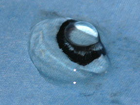 lens and vitreous body 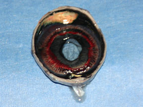 ciliary body and iris viewed from behind | |
The retina contains the sensory
receptors for vision. It is a complex, multilayered structure
which lines most of the rear portion of the eyeball. Its receptors
are rods, which are the receptors for dim light vision, and
the cones which perceive better detail and color than the
rods.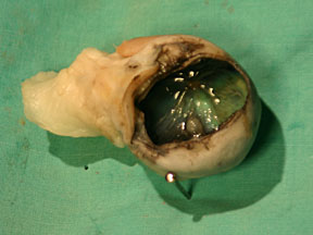 retina and optic disk 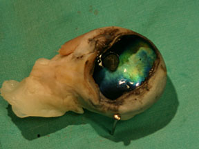 optic disk and tapetum with retina removed 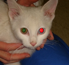 reflection from tapetum (right eye) and blood vessels (left eye) |
The conjunctiva is a thin, transparent membrane that lines the eyelids and covers the front part of the eyeball.
The eyelids are upper and lower folds of skin, lined by conjunctiva that cover and protect the eye. The third eyelid of common domestic animals is a plate of cartilage with some glandular tissue that is covered by conjunctiva and is located medially between the eyelids and the eyeball.
The lacrimal apparatus is the tear producing and draining system. Tears are produced in the lacrimal gland which is located dorsal and lateral to the eyeball up inside the boney orbit that houses and protects the eyeball. The tears wash down over the eye aided by blinking of the eyelids. They are drained from the eye by the lacrimal puncta - 2 small openings in the lid margins located near the medial convergence of the eyelids - the medial canthus. From the lacrimal puncta the tears pass down to the lacrimal sac and then down to the nasal cavity through the nasolacrimal ducts.